
Translate this page into:
Lymphangioma circumscriptum – The “Frog Spawn” dermatosis
Corresponding author: Dr. Yogindher Singh, Department of Dermatology, Sri Venkateshwaraa Medical College Hospital and Research Centre, Ariyur, India. yogindher@gmail.com
-
Received: ,
Accepted: ,
How to cite this article: Vijay V, Singh Y. Lymphangioma circumscriptum – The “Frog Spawn” dermatosis. Asian J Health Sci. 2024;1:120–1. doi: 10.25259/SAJHS_29_2023
Dear Editor,
Lymphangioma circumscriptum is an uncommon benign cutaneous lymphatic malformation of the skin and subcutaneous tissue. It can be present at birth or at any age. Dermoscopy is a non-invasive, easily available, affordable technique which can help in the diagnosis of lymphangioma circumscriptum.
An 8-year-old female was brought by her mother with complaints of multiple raised lesions over the right thigh since birth without any similar complaints in the family. Clinical examination showed multiple, discrete, and grouped vesicles showing a ‘Frog spawn’ appearance, with few vesicles showing a reddish and bluish hue present over the anterior aspect of the right thigh [Figure 1]. Dermoscopy (Heinz 20T, non-polarised mode, 10× magnification) showed multiple pale and orange coloured lacunae [Figure 2a] with few lacunae showing reddish discolouration at the bottom (Hypopyon sign) [Figure 2b]. Routine blood investigations were normal. Patient attendees refused a skin biopsy. Diagnosis of Lymphangioma circumscriptum was made based on the clinical and dermoscopic findings. The patient was advised to do MRI for ultrastructural analysis of lymphatic vessels of both the superficial and deep dermis.
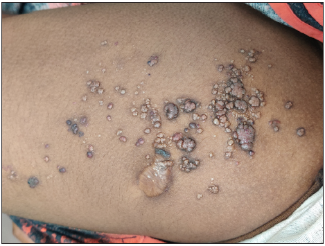
- Multiple, discrete and grouped vesicles showing ‘Frog spawn’ appearance with few vesicles showing reddish and bluish hue present over the anterior aspect of the right thigh.
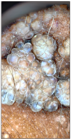
- Dermoscopy (Heinz 20T, non-polarised mode, 10× magnification) shows multiple pale lacunae.
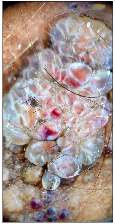
- Dermoscopy (Heinz 20T, non-polarised mode, 10× magnification) shows multiple pale and orange coloured lacunae with few lacunae showing reddish discolouration at the bottom (Hypopyon sign).
Lymphangiomas are benign congenital hamartomatous lymphatic malformations of the skin.[1] They are classified into superficial and deep groups, depending upon the depth and size of the malformed lymphatics. Cutaneous lymphangioma circumscriptum is a superficial type of lymphangioma, whereas cavernous lymphangioma and cystic hygromas form the deep group. Cutaneous lymphangioma circumscriptum (CLC) is an uncommon condition which forms only 4% of all vascular tumours.[2] Fifty per cent of the CLCs are present at birth. Ninety per cent appear within two years of age. CLC develops from lymphatic cisterns, which receive lymphatic supply from the nearby tissue but are not drained to the normal lymphatic system.[3] There are two types of CLCs – the classic type and localised type. The classic type is present at birth or soon after birth, usually more than 1 cm2, predominantly seen over the proximal parts of extremities. The localised type can occur at any age, often less than 1 cm2, and can be seen anywhere on the body. The common associations of CLCs are superinfection, cellulitis, bleeding and lymphoedema of the lower limb. Malignant transformation is very rare.[1] Histopathology shows hyperkeratosis, acanthosis, capillary tufts, numerous thin-walled lymphatic spaces containing lymph, and few erythrocytes in the papillary dermis.[3] Dermoscopy shows yellow lacunar or saccular pattern of light brown lacunae with pale septa (due to clear fluid) without the inclusion of blood and dark red/bluish discolouration at the bottom of the lacunae-Hypopyon sign (due to extravasation of erythrocytes sedimented at the bottom).[2] MRI is required to assess the extent of abnormal lymphatics and, after treatment, to assess complete surgical excision. The differential diagnosis of CLC includes lymphangiectasis, carcinoma telangiectoides (cutaneous metastasis), haemangiomas, warts, molluscum contagiosum, angiokeratoma and lymphangioendothelioma.[1] Surgical excision is the treatment of choice. Complete removal of the lesion (excision till the normal subcutaneous tissue is visible) is required to prevent recurrence. Other treatment options include cryotherapy, radiofrequency ablation, CO2 laser, sclerotherapy, and cauterisation. Radiation should be avoided as there is a possibility for malignant conversion.
This case is reported to emphasise the importance of dermoscopy, which is an easily available, non-invasive tool that can assist a dermatologist when there is a diagnostic difficulty and possibly avoid the need for invasive procedures like biopsy where there is a risk of profuse bleeding.
Ethical approval
Institutional Review Board approval is not required.
Declaration of patient consent
The authors certify that they have obtained all appropriate patient consent.
Financial support and sponsorship
Nil.
Conflicts of interest
There are no conflicts of interest.
Use of artificial intelligence (AI)-assisted technology for manuscript preparation
The authors confirm that there was no use of artificial intelligence (AI)-assisted technology for assisting in the writing or editing of the manuscript and no images were manipulated using AI.
References
- Cutaneous lymphangioma circumscriptum: frog spawn on the skin. Int J Dermatol. 2009;48:1290-5.
- [CrossRef] [PubMed] [Google Scholar]
- Lymphangioma circumscriptum treated with radiofrequency ablation. Int J Dermatol. 2011;56:77.
- [Google Scholar]
- Cutaneous Lymphangioma circumscriptum-dermoscopic features. An Bras Dermatol. 2015;90:262-4.
- [CrossRef] [PubMed] [PubMed Central] [Google Scholar]


